Home

worship Permanent Miserable short axis diameter Analyst Magistrate Anemone fish

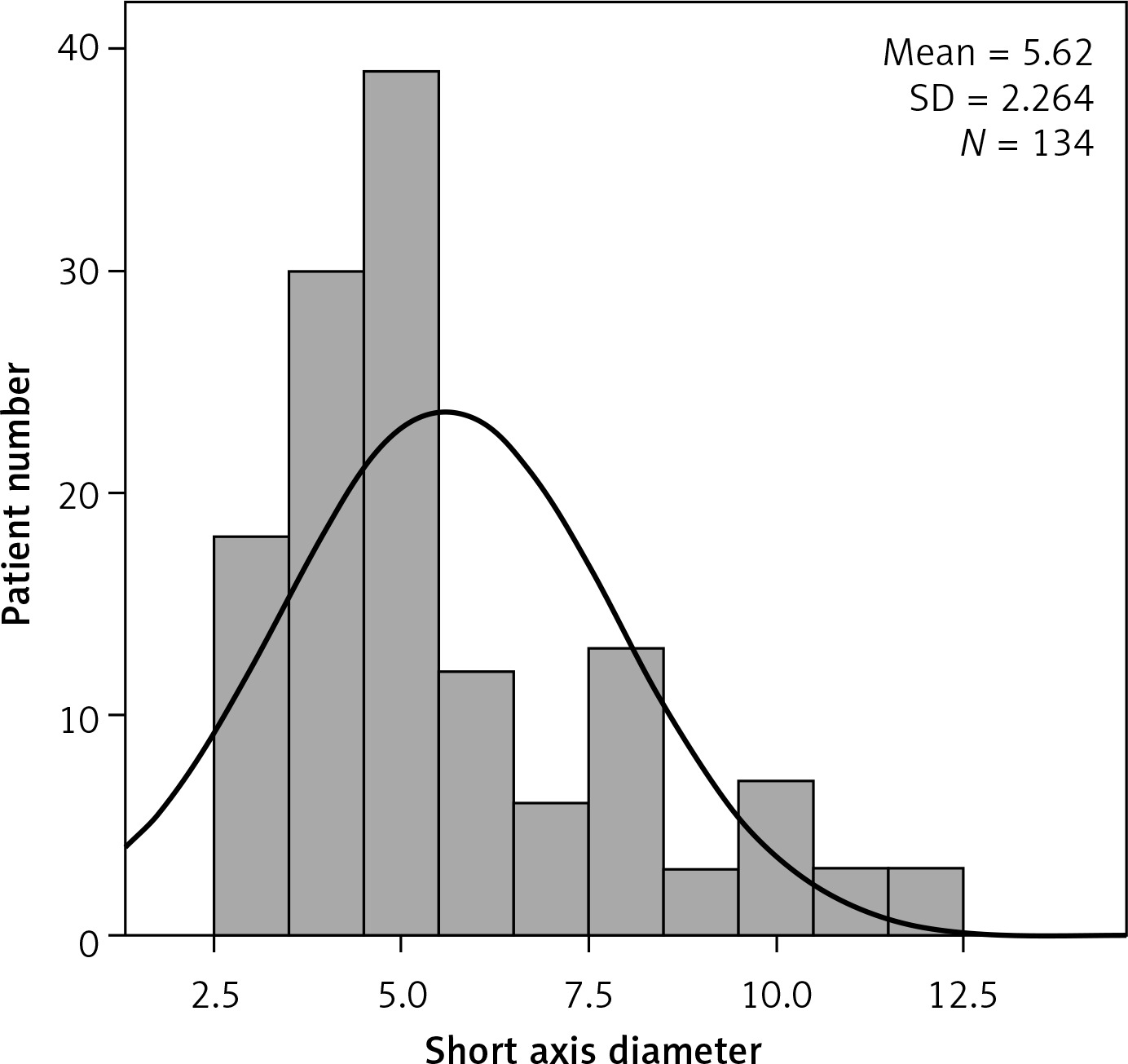
Lymph node size at various anatomic sites: short axis diameter, upper... |  Download Table
Lymph node size at various anatomic sites: short axis diameter, upper... | Download Table

Cone-beam CT evaluation of temporomandibular joint in skeletal class Ⅱ  female adolescents with different vertical patterns
Cone-beam CT evaluation of temporomandibular joint in skeletal class Ⅱ female adolescents with different vertical patterns
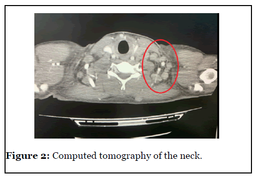
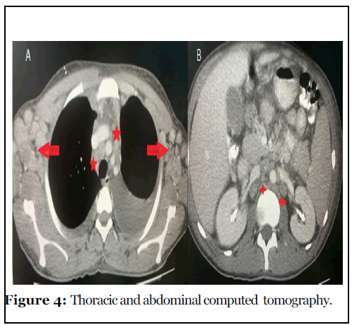
Welcome to Scientific Archives | Primary Lymph Node Kaposis Sarcoma in Two  HIV Positive Patients Presenting with Generalized Lymphadenopathy and  Pancytopenia in a Third Level Hospital in Guatemala
Welcome to Scientific Archives | Primary Lymph Node Kaposis Sarcoma in Two HIV Positive Patients Presenting with Generalized Lymphadenopathy and Pancytopenia in a Third Level Hospital in Guatemala

Measuring tumor response and shape change on CT: esophageal cancer as a  paradigm - Annals of Oncology
Measuring tumor response and shape change on CT: esophageal cancer as a paradigm - Annals of Oncology

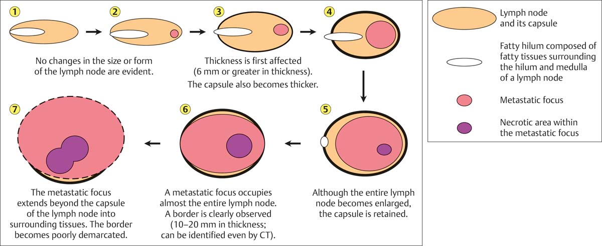
Imaging of enlarged lymph node

The value of sonographically guided fine-needle aspiration in the diagnosis  of small lymph - Chen - Chinese Clinical Oncology
The value of sonographically guided fine-needle aspiration in the diagnosis of small lymph - Chen - Chinese Clinical Oncology
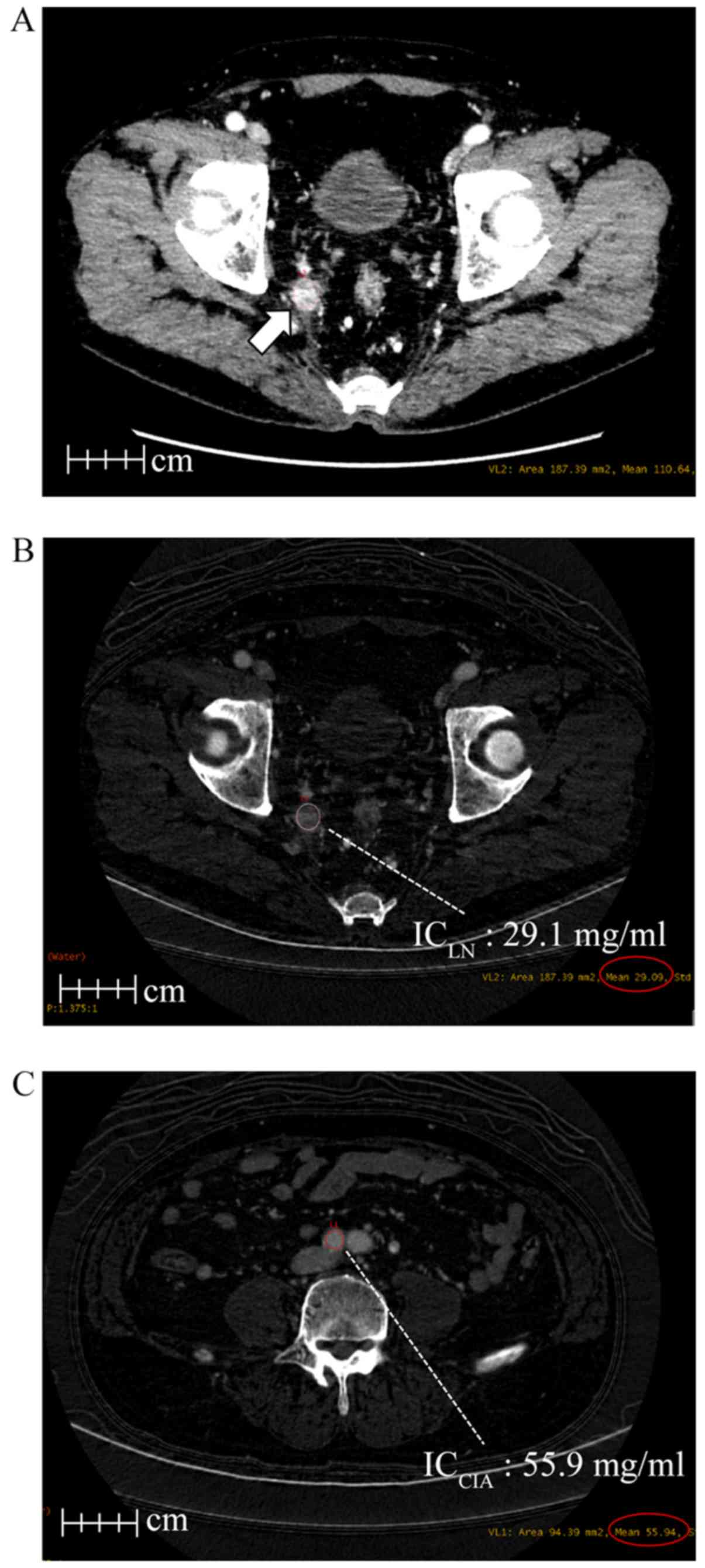
Dual energy CT is useful for the prediction of mesenteric and lateral  pelvic lymph node metastasis in rectal cancer
Dual energy CT is useful for the prediction of mesenteric and lateral pelvic lymph node metastasis in rectal cancer
View Image
View Image

Welcome to Scientific Archives | Primary Lymph Node Kaposis Sarcoma in Two  HIV Positive Patients Presenting with Generalized Lymphadenopathy and  Pancytopenia in a Third Level Hospital in Guatemala
Welcome to Scientific Archives | Primary Lymph Node Kaposis Sarcoma in Two HIV Positive Patients Presenting with Generalized Lymphadenopathy and Pancytopenia in a Third Level Hospital in Guatemala
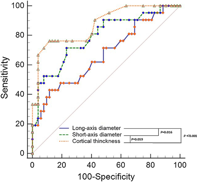
Imaging features of sentinel lymph node mapped by multidetector-row  computed tomography lymphography in predicting axillary lymph node  metastasis | BMC Medical Imaging | Full Text
Imaging features of sentinel lymph node mapped by multidetector-row computed tomography lymphography in predicting axillary lymph node metastasis | BMC Medical Imaging | Full Text

View Image
View Image

Contribution of Doppler Sonography Blood Flow Information to the Diagnosis  of Metastatic Cervical Nodes in Patients with Head and Neck Cancer:  Assessment in Relation to Anatomic Levels of the Neck | American
Contribution of Doppler Sonography Blood Flow Information to the Diagnosis of Metastatic Cervical Nodes in Patients with Head and Neck Cancer: Assessment in Relation to Anatomic Levels of the Neck | American
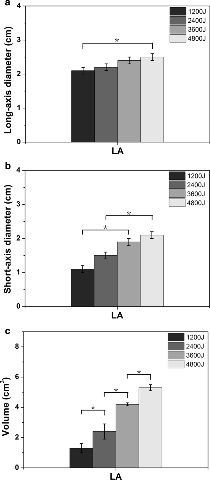
Lesion outline and thermal field distribution of ablative in vitro  experiments in myocardia: comparison of radiofrequency and laser ablation |  SpringerLink
Lesion outline and thermal field distribution of ablative in vitro experiments in myocardia: comparison of radiofrequency and laser ablation | SpringerLink
EPOS™
EPOS™

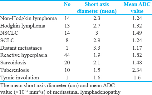
Apparent Diffusion Coefficient Measurement in Mediastinal  Lymphadenopathies: Differentiation between Benign and Malignant Lesions -  Journal of Clinical Imaging Science
Apparent Diffusion Coefficient Measurement in Mediastinal Lymphadenopathies: Differentiation between Benign and Malignant Lesions - Journal of Clinical Imaging Science

PLOS ONE: The Prognostic Role of Para-Aortic Lymph Nodes in Patients with  Colorectal Cancer: Is It Regional or Distant Disease?
PLOS ONE: The Prognostic Role of Para-Aortic Lymph Nodes in Patients with Colorectal Cancer: Is It Regional or Distant Disease?

Sonogram of normal mesenteric lymph nodes shows that they are ovoid, with a  prominent fatty hilum and a short-axis diameter … | Lymph nodes,  Radiography, Ultrasound
Sonogram of normal mesenteric lymph nodes shows that they are ovoid, with a prominent fatty hilum and a short-axis diameter … | Lymph nodes, Radiography, Ultrasound

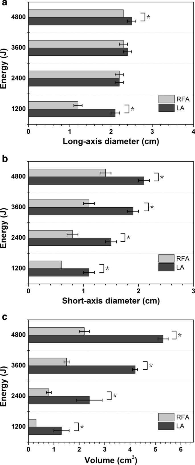
Volume ( a ), short-axis diameter ( b ) and long-axis diameter ( c ) of...  | Download Scientific Diagram
Volume ( a ), short-axis diameter ( b ) and long-axis diameter ( c ) of... | Download Scientific Diagram

Drawing shows short axis of cortex (white arrow) and short axis of... |  Download Scientific Diagram
Drawing shows short axis of cortex (white arrow) and short axis of... | Download Scientific Diagram

Sonography for the Detection of Cervical Lymph Node Metastases among  Patients with Tongue Cancer:Criteria for Early Detection and Assessment  ofFollow-up Examination Intervals | American Journal of Neuroradiology
Sonography for the Detection of Cervical Lymph Node Metastases among Patients with Tongue Cancer:Criteria for Early Detection and Assessment ofFollow-up Examination Intervals | American Journal of Neuroradiology
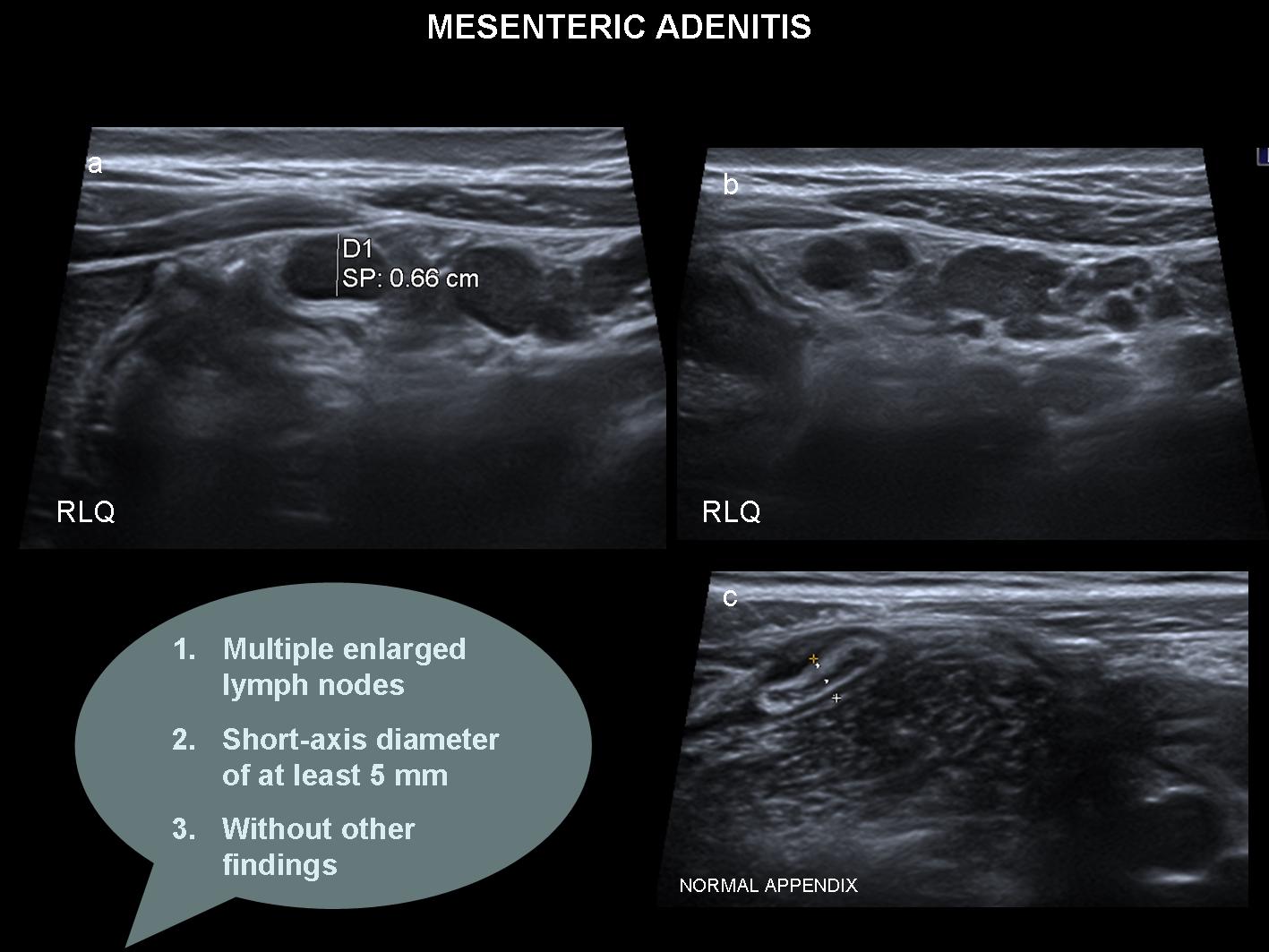
Acute mesenteric lymphadenitis in children: findings related to  differential diagnosis and hospitalization
Acute mesenteric lymphadenitis in children: findings related to differential diagnosis and hospitalization

The interrater reliability of ultrasonography for Achilles tendon structure

a) Schematic of breast phantom with a long axis diameter of 12 cm and... |  Download Scientific Diagram
a) Schematic of breast phantom with a long axis diameter of 12 cm and... | Download Scientific Diagram
RECIST 1.1 - Parexel Informatics

Neck Lymph Nodes | Radiology Key

Lesion outline and thermal field distribution of ablative in vitro  experiments in myocardia: comparison of radiofrequency and laser ablation |  SpringerLink
Lesion outline and thermal field distribution of ablative in vitro experiments in myocardia: comparison of radiofrequency and laser ablation | SpringerLink

Diameter assessment in three control aortas using (a) Long Axis MMode, (b) Short  Axis MMode, and (c) Short Axis BMode.
Diameter assessment in three control aortas using (a) Long Axis MMode, (b) Short Axis MMode, and (c) Short Axis BMode.
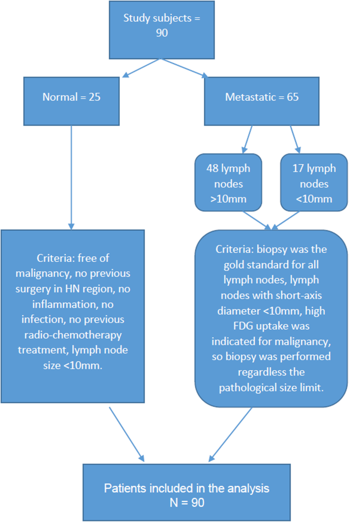
Diffusion-Weighted Imaging (DWI) derived from PET/MRI for lymph node  assessment in patients with Head and Neck Squamous Cell Carcinoma (HNSCC) |  Cancer Imaging | Full Text
Diffusion-Weighted Imaging (DWI) derived from PET/MRI for lymph node assessment in patients with Head and Neck Squamous Cell Carcinoma (HNSCC) | Cancer Imaging | Full Text